Be first to read the latest tech news, Industry Leader's Insights, and CIO interviews of medium and large enterprises exclusively from Healthcare Business Review
Thank you for Subscribing to Healthcare Business Review Weekly Brief
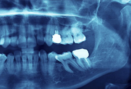
Medical Imaging becomes more Accurate and Easier with 3D X-Ray Techniques
The researchers may use the new approach to measure a 3D image before the sample can be destroyed which may be useful for delicate biological samples.
By
Healthcare Business Review | Thursday, January 14, 2021
Stay ahead of the industry with exclusive feature stories on the top companies, expert insights and the latest news delivered straight to your inbox. Subscribe today.
Today’s healthcare professionals have access to increasingly accurate images with which they can screen, diagnose, and guide patients through the advent of every modern imaging techniques–combined tomography, magnetic resonance imaging, ultrasound, and digital imaging. Medical images are a special type of images which can be used to diagnose patients’ diseases. There are several ways of obtaining these images, such as CT-scan, MRI, and X-ray imaging. Such types of imaging methods usually lead to poor contrast medical images which prevent the doctor from observing and could result in a wrong diagnosis. Using enhanced imaging techniques is essential.
An international research team has discovered two new approaches to 3D images created using X-rays that could enhance disease screening, studied rapid processes, analyze material properties, and offer structural information on opaque objects with unique details. X-rays pass through materials which cannot pass through visible light because of high energy and short wavelength. In many applications, however, the use of 3D X-ray imaging technique is difficult as long-term exposure to x-rays is necessary. One approach may allow 3D imaging of sensitive biological sample or studying very speedy processes, to speed up the development of more durable materials.