Thank you for Subscribing to Healthcare Business Review Weekly Brief

Innovations Changing Landscape of Cardiovascular Tomography and Imaging
Healthcare Business Review
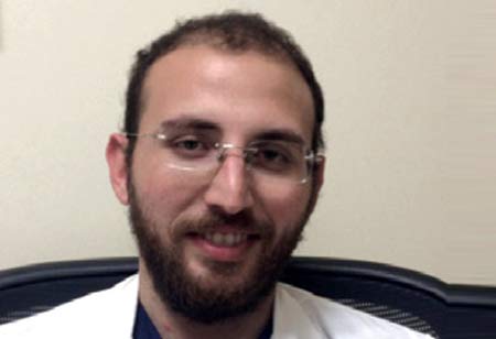
With extensive experience and expertise in Cardiovascular Tomography and imaging, Giuseppe Muscogiuri is currently acting as the Director of Cardiovascular Radiology at Instituoauxologico. He is responsible for conducting studies focused on coronary artery disease, vascular pathology and advanced cardiovascular imaging for CT and MRI.
PLEASE TELL OUR READERS ABOUT YOUR JOURNEY IN THE INDUSTRY.
My journey in the industry has been nothing short of a roller coaster ride. I spent time at Centro Cardiologico Monzino in Milan and many other esteemed institutions during my training. I also worked at the Medical University of South Carolina and Sapienza Università di Roma, Rome. Gathering insights and experiences over time, I was visiting scholar at KU Leuven, Belgium.
Currently, I am the acting director of Cardiovascular Radiology at Institutoauxologico, where I look over advanced imaging for cardiovascular CT and MRI. My focus, mainly, is coronary artery disease and vascular pathology.
WHAT ARE THE RECENT TRENDS IN THE CARDIOVASCULAR IMAGING SPACE?
We have taken leaps and bounds in the technological aspect, which allows us to evaluate plaque composition, especially the quantification of plaque. As a result, we can have a personalized approach with a quantitative evaluation of volumes, flows and tissue characterization. We can use these quantitative techniques to have a specific pathway for each patient with cardiovascular disease. So the future will be a very personalized technique,
COULD YOU TALK ABOUT THE IMPORTANCE OF COMPUTERIZED TOMOGRAPHY.
The real advantage of Cardiac CT is the possibility of evaluating coronary artery disease non-invasively. This means you can determine plaque and coronary plaque that cause coronary artery stenosis. You can also assess the Irish plaque, which can cause a myocardial infarction, an acute event.
The other interesting thing is that with cardiac CT, you can also evaluate the presence of ischemia. So if you perform a dedicated acquisition called CT myocardial perfusion, you can also evaluate the presence of ischemia. These plaques can cause myocarditis ischemia. So by a CCT arrest, computed tomography angiography, and with some dedicated software, the FFRCT, you can evaluate the non-invasively FFR. Furthermore, CT can also be useful for the evaluation of vascular diseases. They can evaluate the valve and are extremely important for the procedure for the planning of the surgical approach.
PLEASE TELL OUR READERS ABOUT PROJECT INITIATIVES THAT YOU ARE CURRENTLY WORKING ON.
We are working on a project which is a multicenter study involving different sites across Europe and US. And we are working on the evaluation of CT perfusion and also the characterization of plaque. So my research activities now are very focused on both myocardial perfusion and characterization of plaque with the iris grafter.
With the advances in Artificial Intelligence, our lives are also changing. Artificial intelligence has the prospect of speeding up the clinician’s work and increasing the diagnosis’s precision
HOW DO YOU ENVISION THE FUTURE?
With new technologies, the cardiovascular imaging space is heading to precision medicine. We can provide a personalized approach for each patient. In the future, we will be using a non-invasive technique with a low radiation dose of contrast agents. We will be able to provide precise information for each patient.
With the advances in Artificial Intelligence, our lives are also changing. Artificial intelligence has the prospect of speeding up the clinician’s work and increasing the diagnosis’s precision. Using algorithms that can speed up the time of acquisition and reporting can help the clinician have a better prognostic stratification.


